ankle/foot pain

Clinical Pattern Recognition- Ankle & Foot Pain

From anatomy to discovering the patient!

Ankle foot pain.jpg
  • Ankle joint accounts for 10% to 34% of all sport-related injuries, with lateral ankle sprain comprising 77% to 83% of these injuries
  • The overall incidence of lateral ankle sprain may be underestimated because approximately 50% do not seek medical attention after injury
  • 100% of 117 therapists who responded noted plantar fasciitis was the most common foot condition seen in the clinic
  • In the athletic population, plantar fasciitis is a common injury reported by both high school, competitive and recreational distance runners

See more prevalence information in the Clinical Pattern Recognition: Orthopaedics app here

Meet the 8 common ankle/foot pain patients from the Achilles Tendonitis Clinical Practice Guidelines, Ankle Stability and Movement Coordination Clinical Practice Guidelines, Heel Pain Clinical Practice Guidelines, and MORE!


Clinical Pattern Recognition

Click on the pain pattern to learn about the patients and develop your clinical patterns!

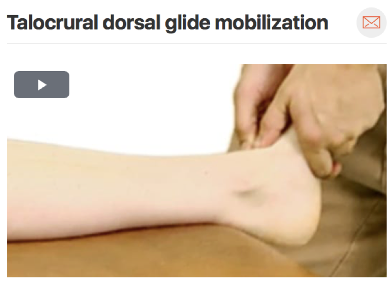
2. Ankle arthrosis- Ankle pain and mobility deficits (Watch 1 min video)

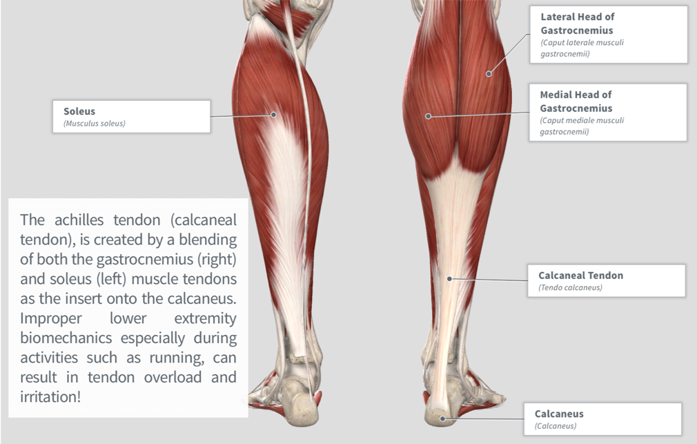
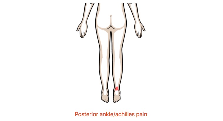
2. Achilles tendinosis- Ankle stiffness and muscle power deficits (Watch 1 min video | Step-by-step Guide)

3. Tibialis anterior tendonitis/Medial tibial stress syndrome- Leg pain and muscle power deficits (Watch 1 minute video)

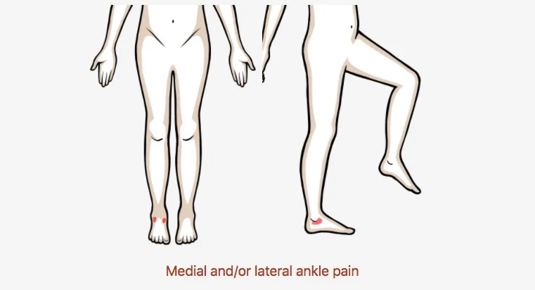
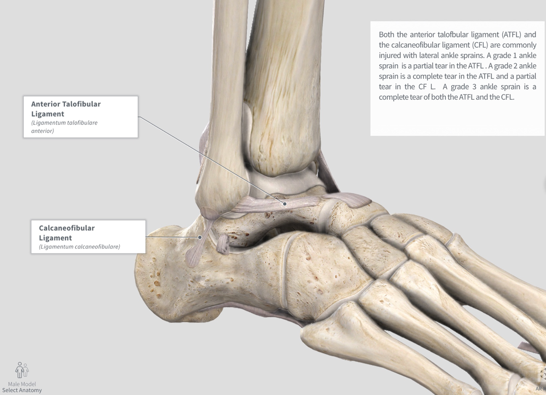
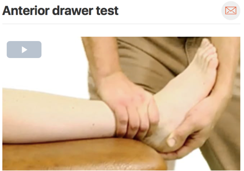
4.  Ankle sprain- Ankle stability and movement coordination impairments (Watch 1 min video | Step-by-Step Guide)

5. Hallux rigidus- Great toe pain and mobility deficits (Watch 1 min video)

6. Posterior tibialis tendinitis/tendinosis- Ankle pain and muscle power deficits (Watch 1 min video | Step-by-Step Guide)

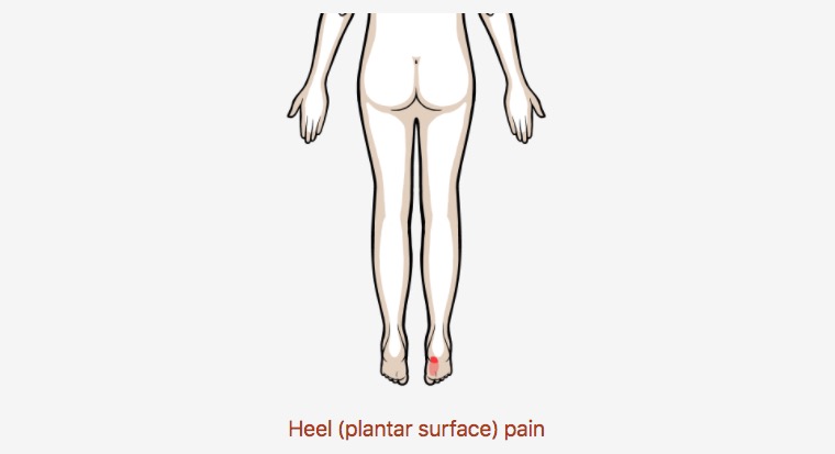
7. Plantar fasciitis - Heel pain (Watch 1 min video | Step-by-Step Guide)

8. Tarsal tunnel syndrome- Ankle and foot radiating pain (Watch 1 min video)

Achilles Tendinopathy

Muscle Power Deficits

Posterior ankle/achilles pain

Patients with varying degrees of achilles tendon pathology making presentation and potential treatment differ slightly; however, they will usually present with fairly localized pain in the achilles tendon and toward the heel!

For more clinical findings click here!

Anatomy

Image via Complete Anatomy by 3D4 Medical

Image via Complete Anatomy by 3D4 Medical

Special Tests

The arc sign is and excellent assessment to help rule in presence of achilles tendon pathology due to it’s high specificity rating!(Click image to watch 1-2 minute video)

The arc sign is and excellent assessment to help rule in presence of achilles tendon pathology due to it’s high specificity rating!

(Click image to watch 1-2 minute video)

Treatment

Insufficient dorsiflexion can be a contributing factor to achilles irritation. If this motion is limited, the achilles can experience increased loading eventually leading to tissue disruption. The gastrocnemius muscle can be a source of movement res…

Insufficient dorsiflexion can be a contributing factor to achilles irritation. If this motion is limited, the achilles can experience increased loading eventually leading to tissue disruption. The gastrocnemius muscle can be a source of movement restriction and should be addressed! (Soft tissue mobilization can be executed in this position as well)

(Click image to watch 1-2 minute video)

Therapeutic Exercise

The gastrocnemius (also achilles tendon) is subject to the highest (eccentric) torque demand during gait, which is further increased while running! As other impairments are being addressed, you may want to consider improving eccentric loading capaci…

The gastrocnemius (also achilles tendon) is subject to the highest (eccentric) torque demand during gait, which is further increased while running! As other impairments are being addressed, you may want to consider improving eccentric loading capacity. (Click image to watch 1-2 minute video)

Posterior Tibialis Tendinopathy

Muscle Power Deficits

Medial ankle and foot (diffuse) pain

Patients with posterior tibialis tendinopathy typically present to clinic with diffuse medial ankle/foot pain often with insidious onset. In more severe cases pain can also radiate into the medial calf region!

For more clinical findings click here!

Anatomy

Image via Complete Anatomy by 3D4 Medical

Image via Complete Anatomy by 3D4 Medical

Midfoot Mobility

Remember, during patient assessment, it is imperative to examine up and down the kinetic chain in order to gain a more well-rounded understanding of the patient’s pathology. That being said, mid foot mobility is still something that should be addres…

Remember, during patient assessment, it is imperative to examine up and down the kinetic chain in order to gain a more well-rounded understanding of the patient’s pathology. That being said, mid foot mobility is still something that should be addressed!

(Click image to watch 1-2 minute video)

Posterior Tibialis Assessment

In order to better rule in the possibility of posterior tibialis tendinopathy, the therapist should elongate, contract, and palpate the muscle belly and tendon looking for reproduction of symptoms. Here is an example of how to properly elongate!&nbs…

In order to better rule in the possibility of posterior tibialis tendinopathy, the therapist should elongate, contract, and palpate the muscle belly and tendon looking for reproduction of symptoms. Here is an example of how to properly elongate!   (Click image to watch 1-2 minute video)

Treatment

Often, lack of dorsiflexion range of motion can result in compensatory mechanisms through the midfoot contributing to irritation of the posterior tibialis tendon. A high-velocity low amplitude thrust to the talocrural joint can help improve this ran…

Often, lack of dorsiflexion range of motion can result in compensatory mechanisms through the midfoot contributing to irritation of the posterior tibialis tendon. A high-velocity low amplitude thrust to the talocrural joint can help improve this range of motion deficit! (Click image to watch 1-2 minute video)

Therapeutic Exercise

Motor coordination both proximally and distally can contribute to excessive pronation. A squat assessment can also double as a therapeutic exercise by cueing the patient to maintain a neutral mid foot position while performing the movement! (Click i…

Motor coordination both proximally and distally can contribute to excessive pronation. A squat assessment can also double as a therapeutic exercise by cueing the patient to maintain a neutral mid foot position while performing the movement! (Click image to watch 1-2 minute video)

Lateral Ankle Sprains

Ankle Stability and Movement Coordination Impairments

Medial and/or Lateral Ankle Pain

Ankle sprains are a very common injury among the younger and physically active population. It is important to know how to properly rehab an ankle sprain to help prevent another one from reoccurring. Look below for some things to consider!

If you do not know the common clinical findings no problem! Click here


Anatomy

Image via Complete Anatomy 2018 by 3D4 Medical

Image via Complete Anatomy 2018 by 3D4 Medical

Common Movement Fault

For lower extremity injuries it is important to watch how your patient walks, this can tell you a lot about their movement patterns. A common movement fault seen in people who sprain their ankle is a lack of dorsiflexion. While lack of dorsiflexion …

For lower extremity injuries it is important to watch how your patient walks, this can tell you a lot about their movement patterns. A common movement fault seen in people who sprain their ankle is a lack of dorsiflexion. While lack of dorsiflexion is not the only factor that can contribute to a risk of an ankle sprain. If the ankle is stiff and is unable to move into full dorsiflexion it will try and gain that range by taking the path of least resistance. Which could lead someone to roll over their ankle causing a lateral ankle sprain.

Special Tests

The anterior drawer test can be used to assess anterior talofibular ligament integrity. This tendon is commonly injured with lateral ankle sprains. Make sure to assess the uninvolved side before assessing the involved side in order to see what is “n…

The anterior drawer test can be used to assess anterior talofibular ligament integrity. This tendon is commonly injured with lateral ankle sprains. Make sure to assess the uninvolved side before assessing the involved side in order to see what is “normal” for the patient. (Click image to watch 1-2 minute video)

Treatment

After your patient has moved through the acute phase of their injury it is important to gain full range of motion of the ankle. This can be achieved through posterior talocrual mobilizations. Grade I and II mobilizations can be used to reduce pain w…

After your patient has moved through the acute phase of their injury it is important to gain full range of motion of the ankle. This can be achieved through posterior talocrual mobilizations. Grade I and II mobilizations can be used to reduce pain where Grade III and IV mobilizations can be used to help increase ROM.

Therapeutic Exercise

Balance is very important in the treatment of ankle sprains in order to help the ankle regain proprioception and prevent future ankle sprains. (Click image to watch 1-2 minute video)

Balance is very important in the treatment of ankle sprains in order to help the ankle regain proprioception and prevent future ankle sprains. (Click image to watch 1-2 minute video)

Plantar Fasciitis

Heel pain

Heel (plantar surface) pain

Plantar fasciitis is one of the most common foot conditions seen in the clinic. It commonly presents with pain with the first steps in the morning or pain after standing for long periods of time. Look below for some things to consider!

If you do not know the common clinical findings no problem! Click here


Anatomy

      Image via Complete Anatomy 2018 by 3D4 Medical

      Image via Complete Anatomy 2018 by 3D4 Medical

Common Movement Fault

For lower extremity injuries it is important to watch how your patient walks, this can tell you a lot about their movement patterns. A common movement fault seen in people with plantar fasciitis is a lack of dorsiflexion. Force is not evenly distrib…

For lower extremity injuries it is important to watch how your patient walks, this can tell you a lot about their movement patterns. A common movement fault seen in people with plantar fasciitis is a lack of dorsiflexion. Force is not evenly distributed through the foot when the ankle does not move through it’s full range of motion. (Click image to watch 1-2 minute video)

Special Tests

The windlass test is a key test to perform when evaluating someone who you suspect has plantar fasciitis. This test is very specific and can rule in the pathology. Pulling the big toe back puts the plantar fascia on stretch and should reproduce the …

The windlass test is a key test to perform when evaluating someone who you suspect has plantar fasciitis. This test is very specific and can rule in the pathology. Pulling the big toe back puts the plantar fascia on stretch and should reproduce the patients pain. (Click image to watch 1-2 minute video)

Treatment

Gaining full range of motion of the talocrural joint is important to achieve to help distribute the ground reaction force as it moves through the body. This can be achieved through posterior talocrual mobilizations. Grade III and IV mobilizations ca…

Gaining full range of motion of the talocrural joint is important to achieve to help distribute the ground reaction force as it moves through the body. This can be achieved through posterior talocrual mobilizations. Grade III and IV mobilizations can be used to help increase ROM.

Therapeutic Exercise

It is important to have your patient work in gaining dorsiflexion range of motion at home if you are also working on it in the clinic! This is an example of one of many stretches that can be used to help gain dorsiflexion range of motion. (Click ima…

It is important to have your patient work in gaining dorsiflexion range of motion at home if you are also working on it in the clinic! This is an example of one of many stretches that can be used to help gain dorsiflexion range of motion. (Click image to watch 1-2 minute video)