Sports Health

The Fear of Re-injury: Psychosocial Impact on Return to Sport “Do They Think They Are Ready”

Dr. Jonathan Hartman PT, DPT, OCS, CSCS, FAAOMPT

Dr. Marshall LeMoine PT, DPT, OCS, SCS, CSCS, FAAOMPT  

ACL 1.png

It has been known for quite some time that athletic performance is intimately connected to psychosocial views, beliefs, and readiness to perform. It is still rarely held in high regards when compared to physical impairment testing amongst musculoskeletal and performance experts.

GOAL: 

  • Provide tests & measures regarding psychosocial return to sport testing 

  • Ways to to implement these test & measures seamlessly into your practice

IMAGINE:

  • Athletes that you believe were held back from their previous performance levels due to psychological factors…

ACL 2.png

The question is, “was it ever addressed?”

CURRENT:

  • Psychosocial return to sport research is highly focused on outcomes with ACL reconstruction (ALC-R) surgery

  • Purpose of this surgery: 

    • Fix the mechanical failure in the body

    • Retrain the athlete back to full function and sport

ACL 3.png

Why does standardized psychosocial testing become more of a specialized subpopulation?

Many athletes are not returning to or surpassing prior level of function due to possible psychosocial barriers. 

  • These barriers can greatly harm: 

    • Overall surgical outcomes

    • Life enjoyment

    • Postoperative complications & comorbidities  

  • Statistics:

    • 21% of postoperative athletes do not return to sport at all

    • 45% do not return to the same pre-injury level of elite sport participation

    • 40-50% of patients do not even return to recreational sports participation

  • Overall:

    • Post-operative patients get less physical exercise 

    • High rate of post-surgical ACL-R weight gain (3.8x more likely to be overweight in 3-10 years after ACL-R surgery)

    • Lack of return to sports activities with increased weight gain

    • >50% of ACL-R patients will develop increased knee OA at a much faster rate than normal

Evidence: 

In this first study (Lentz et al., 2015), physical impairments [quadriceps index (QI), quadriceps strength/body weight (QSBW), hamstring: quadriceps strength ratio (HQ ratio)], pain intensity, self-report of function (International Knee Documentation Committee (IKDC), and psychosocial (Tampa Scale for Kinesiophobia–shortened form (TSK-11) measurements were collected at 6 months and 1 year after surgery using 73 athletes with ACL Reconstruction. At 1 year the subjects were divided into ‘‘return-to-sport’’ or ‘‘not return-to-sport’’ subgroups based on their self-reported return to preinjury sport status. Patients in the “not return-to-sport” subgroup were subcategorized as NRTS-Fear/Confidence if fear of reinjury/lack of confidence was the primary reason for not returning to sports, and all others were categorized as NRTS-Other

This study showed that there is a strong association between a high TSK-11, a low QSBW, and a low IKDC at 1 year in the NRTS-Fear/Confidence subgroup vs the “return-to-sport” group or the NRTS-Other group. This suggests there is a strong psychologically mediated functional limitation with full post-surgical return to sport. Overall, this study supports a growing body of evidence highlighting the importance of measuring and addressing pain-related fear of reinjury, in addition to physical impairments, in standardized return-to-sport criteria (10). 

ACL 4.png
ACL 5.png

Another study (Arden et al., 2013) looked at 187 recreational and competitive athletes, who were given the psychological readiness to return to sport, fear of re-injury, and sport locus of control subjective questionnaires. They were also asked to estimate of the number of months it would take to return to sport both preoperatively and at 4 months postoperatively. At 12 months post ACL-R only 56 athletes (31%) had returned to their preinjury level of sports participation. The results support that psychological responses before surgery and in the early recovery (4 months) were significantly associated with returning to preinjury level of sport at 12 months. This suggests that attention to psychological recovery should start to be implemented in return to function criteria, and start early, even possibly before surgery. This study concludes that screening for maladaptive psychological thought process’ in athletes prior to and soon after surgery may help clinicians identify those athletes at risk of not returning to their preinjury level of sport by 12 months (5).

This next study (Arden et al., 2014) shows how important psychosocial testing can be even with a population that is participating at a recreational level. In this cross-sectional study 164 subjects completed multiple questionnaires at 1–7 years after ACL-R surgery. The questionnaires included a knee self-efficacy (K-SES), health locus of control (MHLOC-C), psychological readiness of return to sport (ACL-RSI), and a fear of re-injury (TSK). At follow-up only 40% (66/164) had returned to their preinjury activity level. Those who did return had a more positive psychological response, reported better knee function in sport and recreational activities, perceived a higher knee-related quality of life, and were more satisfied with their current knee function. In those that did not return, the main reasons identified were not trusting the surgical knee (28%), fear of a new knee injury (24%), and poor knee function (22%). Psychological readiness to return to sport, measured with the ACL-Return to Sport after Injury scale ACLR-SI was most strongly associated with returning to the preinjury activity. Meaning if the athlete has a lower K-SES and ACL-RSI score and a higher TSK-11 score, then this may lead to a lower return to preinjury activity 1-7 years post surgically. This study suggests that including interventions aimed at improving these aspects in postoperative rehabilitation programs could be warranted to improve the rate of return to sport and recreational activities (3). 

ACL 6.png
ACL 7.png

Next, there is a very pertinent systematic reviews (Te Wierike et al., 2013) which support psychosocial testing as an integral piece regarding return to sport and recreation. This systematic review shows that high scores on the “internal health locus of control” and “self-efficacy” tests were useful cognitive factors to facilitate recovery. Also, this study showed that postoperatively ACL-R athletes with a low level of fear of reinjury had the best knee outcomes when returning to sport. In addition, athletes who returned to sport exhibited less fear of reinjury and were more experienced athletes when compared to athletes who did not return to sport (11).

Finally, a meta-analysis of 25 studies, evaluating over 942 injured competitive athletes, uncovered three core themes across the studies chosen. The first is that emotion is associated with, and connected to, rehabilitation outcomes. The second is that there is importance in cognitions associated with rehabilitation outcomes. Third is that those cognitions correlate to behaviors associated with rehabilitation outcomes. These studies support that injury and performance-related fears, anxiety, and confidence were the most common psychosocial factors associated with rehabilitation outcomes. Therefore, practitioners need to recognize that an injured athlete’s thought, feelings, and actions may very well influence the outcomes of rehabilitation, as well as readiness to return to play. 

It can be seen that progressing our profession forward will involve a deep understanding of athletes biopsychosocial connection, and our ability to tease out which is the most limiting factor associated to suboptimal return to function/sport. Our current interpretation of successful rehab may be overly simplified, and practitioners must ensure that injured athletes are physically and psychologically ready to return to sport, with an increased emphasis in non-physical impairments. Practitioners should not assume that physical and psychosocial recovery from injury occurs within the same timeframe or that they are both improving with increased function. They need to be separately and specifically tested (9). As we head into an era focused on true biopsychosocial treatment and individualized sport specific progressive care, it is evident that a holistic approach to patient’s recovery should be addressed to attain the best possible outcomes.

ACL 8.png

Research Quick Reference:

Practitioners need to ensure injured athletes are physically, psychologically, socially, tactically, and technically ready to return to sport (Forsdyke 2016)

Use for Pre-op to 4 months to predict who will return to sports at 12 months (Ardern 2013): 

  • TSK-11, 

  • ISP

  • ERAIQ

  • SRLC 

Use for 6 months & 12 months to predict who will be psychologically ready to return to sport (Lentz 2014):

  • QSBW

  • TSK-11

  • IKDC 

Not returning to sport reasons (Ardern 2014): 

  • Not trusting the knee (28%)

  • Fear of a new injury (24%)

  • Poor knee function (22%)

Use for not returning to sport (Ardern 2014):

  • ACL-RSI

  • K-SES

  • TASK-11 

An athlete with a lower K-SES score, lower ACL-RSI score, and a higher TSK-11 score will have a lower return rate to preinjury physical activity (Arden 2014)

Psychosocial Subjective: 

  • ACL -RSI  score >42 for practice,  score >56 for play (Ardern 13’, Webster 18’)

  • TSK-11 score <17  for practice , score <15 play (Lentz 2015, Woby 2005)

Knee function Subjective: 

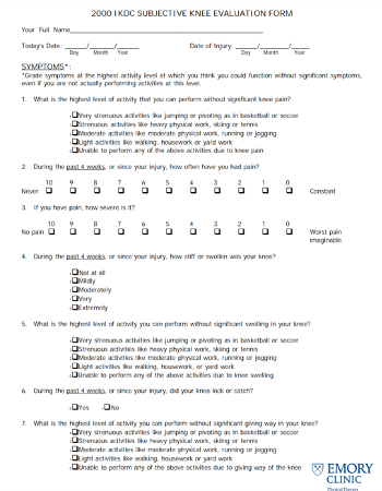
  • IKDC Question #10, score >9 (M.O.O.N. Group Guidelines)

Citations

  1. Ardern, C. L., Taylor, N. F., Feller, J. A., & Webster, K. E. (2014). Fifty-five per cent return to competitive sport following anterior cruciate ligament reconstruction surgery: an updated systematic review and meta-analysis including aspects of physical functioning and contextual factors. British Journal of Sports Medicine. Nov2014, 48(21), 1543. doi:10.1136/ bjsports-2013-093398

  2. Ardern, C. L., Taylor, N. F., Feller, J. A., Whitehead, T. S., & Webster, K. E. (2015). Sports Participation 2 Years After Anterior Cruciate Ligament Reconstruction in Athletes Who Had Not Returned to Sport at 1 Year: A Prospective Follow-up of Physical Function and Psychological Factors in 122 Athletes. American Journal of Sports Medicine (AM J SPORTS MED), Apr2015. doi:http://0-dx.doi.org.catalog.llu.edu/10.1177/0363546514563282 

  3. Ardern, C. L., Osterberg, A., Tagesson, S., Gauffin, H., Webster, K. E., Kvist, J., & Österberg, A. (2014). The impact of psychological readiness to return to sport and recreational activities after anterior cruciate ligament reconstruction. British Journal of Sports Medicine (BR J SPORTS MED), Dec2014. doi:http://0-dx.doi.org.catalog.llu.edu/10.1136/ bjsports-2014-093842 

  4. Ardern CL, Webster KE, Taylor NF, Feller JA. Return to sport following anterior cruciate ligament reconstruction surgery: A systematic review and meta-analysis of the state of play. Br J Sports Med 2011;45(7):596-606.

  5. Arden CL, Taylor NF, Feller JA, Whitehead TS, Webster KE. Psychological Responses Matter in Returning to Preinjury Level of Sport After Anterior Cruciate Ligament Reconstruction Surgery. The American Journal of Sports Medicine. 2013; 41 (7): 1549-1558. Doi:10.1177/0363546513489284.

  6. Chmielewski TL, Zeppieri G Jr, Lentz TA, et al. Longitudinal changes in psychosocial factors and their association with knee pain and function after anterior cruciate ligament reconstruction. Phys Ther 2011; 91: 1355–1366.

  7. Everhart, J. S., Best, T. M., & Flanigan, D. C. (2015). Psychological predictors of anterior cruciate ligament reconstruction outcomes: a systematic review. Knee Surgery, Sports Traumatology, Arthroscopy (KNEE SURG SPORTS TRAUMATOL ARTHROSC), Mar2015. doi:http://0-dx.doi.org.catalog.llu.edu/10.1007/s00167-013-2699-1

  8. Flanigan, D. C., Everhart, J. S., Pedroza, A., Smith, T., & Kaeding, C. C. (2013). Fear of reinjury (kinesiophobia) and persistent knee symptoms are common factors for lack of return to sport after anterior cruciate ligament reconstruction. Arthroscopy: The Journal of Arthroscopy & Related Surgery (ARTHROSCOPY), Aug2013. doi:http://0- dx.doi.org.catalog.llu.edu/10.1016/j.arthro.2013.05.015

  9. Forsdyke D, Smith A, Jones M, et al. Psychosocial factors associated with outcomes of sports injury rehabilitation in competitive athletes: a mixed studies systematic review. Br J Sports Med 2016;50:537-544.

  10. Lentz, T. A., Zeppieri, G., George, S. Z., Tillman, S. M., Moser, M. W., Farmer, K. W., & Chmielewski, T. L. (2015). Comparison of Physical Impairment, Functional, and Psychosocial Measures Based on Fear of Reinjury/Lack of Confidence and Return-to-Sport Status After ACL Reconstruction. American Journal of Sports Medicine (AM J SPORTS MED), Feb2015. doi:http://0-dx.doi.org.catalog.llu.edu/10.1177/0363546514559707

  11. Te Wierike, et al. Psychosocial factors influencing the recovery of athletes with anterior cruciate ligament injury: A systematic review. Scandinavian Journal of Medicine & Science in Sports 2013.

  12. Thomee P, Wahrborg P, Borjesson M, et al. Self-efficacy of knee function as a pre- operative predictor of outcome 1 year after anterior cruciate ligament reconstruction. Knee Surg Sports Traumatol Arthrosc 2008; 16: 118–127. 

  13. Tjong VK, Murnaghan ML, Nyhof-Young JM, et al. A qualitative investigation of the decision to return to sport after anterior cruciate ligament reconstruction: to play or not to play. Am J Sports Med 2014; 42: 336–342.

  14. Tripp DA, Stanish W, Ebel-Lam A, et al. Fear of reinjury, negative affect, and catastrophizing predicting return to sport in recreational athletes with anterior cruciate liga- ment injuries at 1 year postsurgery. Rehabil Psychol 2007; 52: 74–81.

  15. Wright RW, Haas AK, Anderson J, et al. Anterior cruciate ligament reconstruction rehabilitation: MOON guidelines. Sports Health 2015; 7: 239–243

Lunging Into Stride Length Wrap up!

Here’s our latest in the Sports Health section from Dr. Jonathan Hartman and Dr. Marshall LeMoine. This topic has been broken out into 3 parts due to its’ length so keep an eye out every Wednesday of this month to stay on top of this great article.

We hope you’ve enjoyed this article thus far! To recap here are the links in case you missed them and need a refresher

For your reference during the movement assessment, here’s the Step Back Barbell Lunge Movement Fault Guide and quick reference cheat sheet. If you’d like to read on further, the research and citations are listed below!

Here’s the Step Back Lunge demonstrated by Dr Hartman himself. (In case you missed this in our first post.)

Step Back Barbell Lunge Movement Fault Guide:

Lateral View:

  • Chin neck angle constant 60-90 (Cue: Hold a tennis ball under chin- DNF, Stick on back/ head, allow gaze to follow body)

  • External auditory meatus over shoulder

  • Thumbs always over the bar (To avoid wrist extension and wrist injury, also allows for elbows up for latissimus engagement)

  • Any bar lunge, the bar will ALWAYS be just below C7 spinous process

  • Elbows back and slightly up (should only see one elbow from the side if they are in line)

  • Abdominal Brace (Breath in and out at the top of lunge, never bottom position, no trunk rotation or lateral flexion)

  • Lumbar spine neutral to start

  • Start the lunge with a hip hinge first then allow a knee break and continue with 1:1 knee and hip flexion rate

  • Front knee pulled out over toes 1-3 (throughout closed chain knee flexion of the front leg)

  • No femoral adduction/IR on either leg when in closed chain (on descend or ascend)

  • Front foot does not have excessive pronation

  • Both feet start hip distance apart or slightly narrower, foot that steps back tracks in a straight line and lands on the ground in the same plane as starting foot position

  • Step back and lower (do not touch knee to ground)

  • Re-rack weight by returning to starting position, Look to see that the bar is at the same height on both sides, Lock bar in, then finally Look to check that it is fully locked in, then step out (Always re-rack weight with 3 “L” Check Procedure Look/Lock/Look then step out)

Posterior View:

  • Cervical neutral without lateral flexion

  • Thumbs always over the bar (To avoid wrist extension and wrist injury, also allows for elbows up for lats engagement)

  • Any bar back lunge, the bar will ALWAYS be below C7 spinous process

  • Elbows back, slightly up, same height (Bar parallel to ground)

  • Abdominal Brace (Breath in and out at the top of lunge, never bottom position, no trunk rotation or lateral flexion)

  • Lumbar spine neutral to start

  • Start the lunge with a hip hinge first then allow a knee break and continue with 1:1 knee and hip flexion rate

  • Front foot neutral eversion / inversion (Toe sign)

  • Front knee pulled out over toes 1-3 (throughout closed chain knee flexion of the front leg)

  • No femoral adduction/IR on either leg when in closed chain (on descend or ascend)

  • Front foot does not have excessive pronation

  • Both feet start hip distance apart or slightly narrower, foot that steps back tracks in a straight line and lands on the ground in the same plane as starting foot position

  • Equal leg window always seen from posterior view throughout motion

  • Step back and lower (do not touch knee to ground)

  • Re-rack weight by returning to starting position, Look to see that the bar is at the same height on both sides, Lock bar in, then finally Look to check that it is fully locked in, then step out (Always re-rack weight with 3 “L” Check Procedure Look/Lock/Look then step out)

Quick Look Movement:

  • Bar starts on the rack level of inferior scapular angle

  • Patient steps under and positions bar, patient feels comfortable with bar weight and is ready

  • Set up Trunk to Floor positioning

  • Chin neck angle 60-90 entire time without lateral flexion

  • Turn on Brace (Tactile cue/one long controlled pursed lip exhale breath or breath held for entire motion is OK)

  • One leg steps back and hip hinge initiation with trunk anterior lean to start movement followed by knee flexion

  • After hip hinge initiation, hip and knee flexion at same rate on front leg

  • Step back and lower (do not touch knee to ground)

  • Step back with foot landing in line with starting position

  • Avoid Femoral adduction/ IR

  • Breath at top and reposition as needed

  • Re-rack weight with 3 “L” Check Procedure Look/Lock/Look then step out

Research Quick Reference:

  • Gluteus maximus and medius are better activated with exercises that have a single stance leg component (1)

  • To Increase the hip extensor impulse and hip extensor EMG of the (G-max and BF) use a forwards trunk lean (Average of 107.9° hip flexion) (7)

  • In order to enhance activation of the superficial core musculature unilateral UE weights should be used over bilateral UE weights (14)

  • Joint loading progression for the hip can begin with Single-leg squat → Reverse lunge → Forward lunge (3)

  • Joint loading progression for the knee and ankle can begin with Reverse lunge → Forward lunge → Single-leg squat (3)

  • Walking lunge + weight held in the contralateral arm to the anterior/ moving leg leads to an increase the gluteus medius and vastus lateralis activation. (16)

CITATIONS

  1. Boren K, Conrey C, Le Coguic J, Paprocki L, Voight M, Robinson TK. Electromyographic analysis of gluteus medius and gluteus maximus during rehabilitation exercise. International Journal of Sports Physical Therapy. 2011;6(3):206-223.

  2. Chowdhury, S., & Kumar, N. (2013). Estimation of forces and moments of lower limb joints from kinematics data and inertial properties of the body by using inverse dynamics technique. Journal of Rehabilitation Robotics, 1(2), 93-98

  3. Comfort P, Jones PA, Smith LC, Herrington L. Joint Kinetics and Kinematics During Common Lower Limb Rehabilitation Exercises. Journal of Athletic Training. 2015;50(10):1011-1018. doi:10.4085/1062-6050-50.9.05.

  4. Contreras, Bret. Force Vector Training (FVT). The Glute Guy, 1 July 2010, Bretcontreras.com/load-vector-training-lvt/.

  5. Dwyer MK, Boudreau SN, Mattacola CG, Uhl TL, Latterman C. Comparision of lower extremity kinematics and hip muscle activation during rehabilitation tasks between sexes. J Athl Train. 2010;45(2):181–190

  6. Ekstrom RA, Donatelli RA, Carp KC. Electromyographic analysis of core trunk, hip, and thigh muscles during 9 rehabilitation exercises. J Orthop Sports Phys Ther. 2007;37(12):754–762.

  7. Farrokhi S, Pollard CD, Souza RB, Chen YJ, Reischl S, Powers CM. Trunk position influences the kinematics, kinetics, and muscle activity of the lead lower extremity during the forward lunge exercise. J Orthop Sports Phys Ther. 2008 Jul;38(7):403-9. doi: 10.2519/jospt.2008.2634. Epub 2008 Apr 15.

  8. Flanagan et al (2003). Lower extremity biomechanics during forward and lateral stepping activities in older adults. Clinical Biomechanics, 18(3), 2 14-22. Roger W. Earle (2005). Essential of personal training. National Strength and Conditioning Association.

  9. Hefzy MS, al Khazim M, Harrison L. Co-activation of the hamstrings and quadriceps during the lunge exercise. Biomed Sci Instrum. 1997;33:360–365.

  10. Khaiyat OA, Norris J. Electromyographic activity of selected trunk, core, and thigh muscles in commonly used exercises for ACL rehabilitation. Journal of Physical Therapy Science. 2018;30(4):642-648. doi:10.1589/jpts.30.642.

  11. N Boudreau, Samantha & Dwyer, Maureen & Mattacola, Carl & Lattermann, Christian & Uhl, Tim & Medina McKeon, Jennifer. (2009). Hip-Muscle Activation During the Lunge, Single-Leg Squat, and Step-Up-and-Over Exercises. Journal of sport rehabilitation. 18. 91-103. 10.1123/jsr.18.1.91.

  12. Riemann BL, Lapinski S, Smith L, Davies G. Biomechanical Analysis of the Anterior Lunge During 4 External-Load Conditions. Journal of Athletic Training. 2012;47(4):372-378.

  13. Riemann, Bryan & Congleton, A & Ward, R & Davies, George. (2013). Biomechanical comparison of forward and lateral lunges at varying step lengths. The Journal of sports medicine and physical fitness. 53. 130-8.

  14. Saeterbakken AH, Fimland MS, Navarsete J, Kroken T, van den Tillaar R (2015) Muscle Activity, and the Association between Core Strength, Core Endurance and Core Stability. J Nov Physiother Phys Rehabil 2(2): 028-034. DOI: 10.17352/2455-5487.000022

  15. Saeterbakken, Atle & Fimland, Marius. (2011). Muscle activity of the core during bilateral, unilateral, seated and standing resistance exercise. European journal of applied physiology. 112. 1671-8. 10.1007/s00421-011-2141-7.

  16. Stastny et al (2015). Does the dumbbell-carrying position change the muscle activity in split squats and walking lunges? Journal of Strength and Conditioning Research, 29(11), 3177-3187. Thomas R. Baechle et al (2013) Essentials of strength training and conditioning. National Strength and Conditioning Association.

  17. Stuart MJ, Meglan DA, Lutz GE, Growney ES, An KN. Comparison of intersegmental tibiofemoral joint forces and muscle activity during various closed kinetic chain exercises. Am J Sports Med. 1996; 24(6):792–799.

Lunging Into Stride Length Part III: Lift Progressions of the Lunge to Optimize Performance

Here’s our latest in the Sports Health section from Dr. Jonathan Hartman and Dr. Marshall LeMoine. This topic has been broken out into 4 parts due to its’ length so keep an eye out every Wednesday of this month to stay on top of this great article.

If you missed the part 1 & 2, here are the links to review: Lunging Into Stride Length Part I:  Introducing the Benefits of a Functional Lunge & Lunging Into Stride Length Part II:  Research Based Evidence of Benefits of the Lunge for Strength and Sport Adaptations

Lift Progression

Using current evidence one possible version of a graded progressive treatment protocol for the lunge could focus on first and foremost the unweighted form of the movement, which is an aspect I feel is constantly rushed through with higher level athletes as most of them have done this type of exercise before. But just because you have done something a thousand times does not mean it was done right. In this respect our focus should be to foster athletic intellectual training in order to improve the athlete’s understanding of the correct body movements during this motion in order to enhance performance. Take the time to explain to them the purpose and the benefits of each form correction, and let them feel what each correction can do so that as the weight and variables increase they can problem solve through form corrections themselves. When training high level athletics the lift intention and athletic optimization go hand in hand as the athlete should have an in-depth understanding of how the lift connects to the exact sport movement as this will allow them to picture how it will transfer over into function and may give the seemingly separate esoteric lift a more meaningful sport specific connection.

Focus from bottom to top during starting stance position, then onto the initiation of the motion as this will set up the entirety of the descent phase. Then shift the focus to the anterior open chain moving leg for the forwards lung or the anterior closed chain stationary leg for the reverse step back lunge as it will accept the majority of the force throughout the descent phase. Finally watch the athlete’s power leg stability and trunk control as they push back up during the ascent phase ending in the starting stance position. The movement guide focuses on the dynamic step back / reverse lunge but it can be applied for any lunge variation. Please use the movement fault guide below to guide the common faults seen with this exercise. Next focus on the athletes closed chain weight accepting foot / arch control and knee positioning using small external cues (such as an anterior mirror with a vertical trunk alignment tape, or by having the patient lunge next to a wall for a lateral trunk or knee alignment cue). Next, progress to a multiplanar lunge if applicable to the athlete’s sport specificity. You can also shift the focus on cueing specific muscles with a TheraBand loop around thighs in order to promote the athlete’s femoral eternal rotation and abduction.

neutral trunk lunge.jpg

We can optimize performance faults of the closed chain limb at the forefoot, knee, and hip with the use of TheraBand by having the athlete put the band under the great toe metatarsal head to a loop around the contralateral hand or by wrapping the band from the great toe metatarsal head coursing laterally over the TCN joint then medially behind the gastrocnemius bellies/ knee / thigh ending in a loop around the contralateral hand. The athlete can also perform this motion with bilateral upper extremity patterning or contralateral upper extremity patterning matching their sport specific motions. Perhaps a goalie or a jumper would use a bilateral upper extremity flexion and extension pattern while pushing off one foot in order to catch a ball or create momentum while a sprinter will use contralateral upper extremity patterning to counterbalance lower extremity push off and for full body force transmission. We can also use higher level resistance bands to further challenge the trunk via trunk wrap used to pull the athletes center of mass out of neutral while the athlete resists this force. We can add instability components to the stance leg via Bosu-Ball, Airex-Pad, sand, or external perturbations. Increasing speed or adding a flight phase as seen with an alternating jumping split lunge can also increase the challenge of the exercise. Another progression I do not see frequently performed but is of extreme importance is incorporating sport related equipment such as holding balls, bats, or racquets, as well as starting the movement to a “start” buzzer or pop noise can greatly enhance the sport specificity with quick reactions. Even such things as putting on skis or boots of the sport can be incorporated as some equipment limits specific lower extremity joint motion and thus performing these exercises in and out of this equipment can change kinematics dramatically. Finally, try performing all of these progressions under systemic fatigue as it will highlight any fatigue related movement faults.  “Practice how you want to perform”

breugers wrap lunge frontal view.jpg
lunge with ball sagittal view 2.jpg
bosu lunge.jpg

As you can see, this progression can be extensive, creative, and individualized per athlete and sport. At any point in this progression, extra weight can be added, if hypertrophy or strength is the goal, via kettlebells, dumbbells, or a barbell as well as with many other methods. Using the above evidence we can see holding the weights in differing hands or positions will vary the athlete’s trunk activation. It is suggested to start with lighter weight and then work upwards within the session or throughout sequential sessions based on the form and function of the previous lift or session.

It is slightly controversial when it comes to adding a lot of external cueing and bands to an athlete performing a strength or hypertrophic lift. There are some schools of thought who think this will take away from the lift, but there is value in targeting movement faults or areas of possible future fault in order to maximize athletic function. Thus we can use band wraps as described above but instead of attaching them to the contralateral hand they can be attached to the squat rack on the contralateral side for lower extremity positioning and cueing. We can also challenge the trunk by use offset weight stacks or by adding hanging weights from the barbell via Monster-bands leading to further weighted instability. We can also further challenge the system as a whole by performing lunges with barbell or dumbbells held extended overhead on the ipsilateral or contralateral upper extremity.

Hopefully this article has shed some light on what research has to say about the lunge, as well as taught you a couple of key ways to challenge, progress and modify the lunge exercise to target key muscles for your athletes. Just remember small movement optimization holds immense value with the athletic population and this can be enhanced through a progressive evidence based sport specific training and rehabilitation program.

Stayed tuned for the final section of Lunging into Stride Strength with a complete recap, movement fault guide and references for your review!

CITATIONS

  1. Boren K, Conrey C, Le Coguic J, Paprocki L, Voight M, Robinson TK. Electromyographic analysis of gluteus medius and gluteus maximus during rehabilitation exercise. International Journal of Sports Physical Therapy. 2011;6(3):206-223.

  2. Chowdhury, S., & Kumar, N. (2013). Estimation of forces and moments of lower limb joints from kinematics data and inertial properties of the body by using inverse dynamics technique. Journal of Rehabilitation Robotics, 1(2), 93-98

  3. Comfort P, Jones PA, Smith LC, Herrington L. Joint Kinetics and Kinematics During Common Lower Limb Rehabilitation Exercises. Journal of Athletic Training. 2015;50(10):1011-1018. doi:10.4085/1062-6050-50.9.05.

  4. Contreras, Bret. Force Vector Training (FVT). The Glute Guy, 1 July 2010, Bretcontreras.com/load-vector-training-lvt/.

  5. Dwyer MK, Boudreau SN, Mattacola CG, Uhl TL, Latterman C. Comparision of lower extremity kinematics and hip muscle activation during rehabilitation tasks between sexes. J Athl Train. 2010;45(2):181–190

  6. Ekstrom RA, Donatelli RA, Carp KC. Electromyographic analysis of core trunk, hip, and thigh muscles during 9 rehabilitation exercises. J Orthop Sports Phys Ther. 2007;37(12):754–762.

  7. Farrokhi S, Pollard CD, Souza RB, Chen YJ, Reischl S, Powers CM. Trunk position influences the kinematics, kinetics, and muscle activity of the lead lower extremity during the forward lunge exercise. J Orthop Sports Phys Ther. 2008 Jul;38(7):403-9. doi: 10.2519/jospt.2008.2634. Epub 2008 Apr 15.

  8. Flanagan et al (2003). Lower extremity biomechanics during forward and lateral stepping activities in older adults. Clinical Biomechanics, 18(3), 2 14-22. Roger W. Earle (2005). Essential of personal training. National Strength and Conditioning Association.

  9. Hefzy MS, al Khazim M, Harrison L. Co-activation of the hamstrings and quadriceps during the lunge exercise. Biomed Sci Instrum. 1997;33:360–365.

  10. Khaiyat OA, Norris J. Electromyographic activity of selected trunk, core, and thigh muscles in commonly used exercises for ACL rehabilitation. Journal of Physical Therapy Science. 2018;30(4):642-648. doi:10.1589/jpts.30.642.

  11. N Boudreau, Samantha & Dwyer, Maureen & Mattacola, Carl & Lattermann, Christian & Uhl, Tim & Medina McKeon, Jennifer. (2009). Hip-Muscle Activation During the Lunge, Single-Leg Squat, and Step-Up-and-Over Exercises. Journal of sport rehabilitation. 18. 91-103. 10.1123/jsr.18.1.91.

  12. Riemann BL, Lapinski S, Smith L, Davies G. Biomechanical Analysis of the Anterior Lunge During 4 External-Load Conditions. Journal of Athletic Training. 2012;47(4):372-378.

  13. Riemann, Bryan & Congleton, A & Ward, R & Davies, George. (2013). Biomechanical comparison of forward and lateral lunges at varying step lengths. The Journal of sports medicine and physical fitness. 53. 130-8.

  14. Saeterbakken AH, Fimland MS, Navarsete J, Kroken T, van den Tillaar R (2015) Muscle Activity, and the Association between Core Strength, Core Endurance and Core Stability. J Nov Physiother Phys Rehabil 2(2): 028-034. DOI: 10.17352/2455-5487.000022

  15. Saeterbakken, Atle & Fimland, Marius. (2011). Muscle activity of the core during bilateral, unilateral, seated and standing resistance exercise. European journal of applied physiology. 112. 1671-8. 10.1007/s00421-011-2141-7.

  16. Stastny et al (2015). Does the dumbbell-carrying position change the muscle activity in split squats and walking lunges? Journal of Strength and Conditioning Research, 29(11), 3177-3187. Thomas R. Baechle et al (2013) Essentials of strength training and conditioning. National Strength and Conditioning Association.

  17. Stuart MJ, Meglan DA, Lutz GE, Growney ES, An KN. Comparison of intersegmental tibiofemoral joint forces and muscle activity during various closed kinetic chain exercises. Am J Sports Med. 1996; 24(6):792–799.

Lunging Into Stride Length Part II:  Research Based Evidence of Benefits of the Lunge for Strength and Sport Adaptations

Here’s our latest in the Sports Health section from Dr. Jonathan Hartman and Dr. Marshall LeMoine. This topic has been broken out into 4 parts due to its’ length so keep an eye out every Wednesday of this month to stay on top of this great article.

If you missed last week’s blog post on the Lunging Into Stride Length Part I:  Introducing the Benefits of a Functional Lunge.

Evidence

This next section is split into 3 parts in order to focus on current evidence surrounding muscle activation, the benefits of single leg stance and core activation, and finally how differing variations of the lunge exercise stack up to each other. Let’s start with a 2011 study that utilized EMG to measure the muscle activity of the gluteus maximus and medius of 26 healthy subjects during 18 different lower extremity exercises. This study showed that of the top 4 exercises for gluteal muscle activity the only stance exercise that proved to be of high-level activation was a single leg stance exercise. This study helps to support the idea that exercises incorporating a single leg stance component, such as the movements seen in the ascent and descent of a step lunge will increase the activation of the gluteus maximus and medius muscles (1).

In 2008, Farrokhi et al. performed a study on 10 healthy individuals to determine how a change in trunk position via a forwards lean, neutral, or erect trunk would influence the lower extremity kinematics and kinetics at the hip, knee, and ankle during a forward lunge. This study also monitored how these trunk variations would influence the muscle activity of the lateral gastrocnemius, vastus lateralis, gluteus maximus and biceps femoris. This study showed that the forward trunk lean lunge resulted in an average of 107.9 degrees of hip flexion, and was found to significantly increase the hip extensor impulse and EMG of the gluteus maximus and biceps femoris when compared to the neutral trunk position. This study can further guide our exercise precision and prescription to target the posterior chain via optimizing the trunk angle of the athlete when performing a lung exercises (7).


Forward Trunk Lean Vs. Erect Trunk Lean

forward trunk lunge.jpg
neutral trunk lunge.jpg

In a 2009 study that took 44 healthy individuals, and had them perform a lunge, single-leg squat, and step-up-and-over exercise, and  recorded EMG of 5 muscles (rectus femoris, dominant and nondominant gluteus medius, adductor longus, and gluteus maximus). This study showed that the rectus femoris, gluteus maximus, and dominant side gluteus medius were activated in a progression from least to greatest during the step up and over, lunge, and single leg squat. Interestingly gluteus medius on the non-dominant leg activation was from least to greatest during the single leg squat, step up and over, and then the lunge. This study supports the idea that the lunge muscle activation differs with the dominant and nondominant leg but in either circumstance this exercise is a viable and possible superior choice option for strength training when compared to the step up and over and the single leg squat (11).

Next, surface EMG was used on twelve active female subjects to compare activation of eight trunk, hip/core, and lower limb muscles (erector spinae, rectus abdominis, gluteus maximus, vastus lateralis, rectus femoris, vastus medialis, biceps femoris, and semitendinosus) during the forward lunge, double leg raise, glute bridge, sit-up, and squat. The neutral trunk forward lunge produced significantly higher activation in the vastus medialis, vastus lateralis, and rectus femoris muscles compared to the other exercises, thus supporting the specificity of muscular adaptations to specific lifts (10).

One important take away from these studies is the fact that we can strengthen the lower extremities and gain muscle activation with a majority of the weight emphasis on a single leg. Since we are strengthening with a bias towards one leg the load on the bar or weight held in the hand will be less than a similar closed chain bilateral lower extremity exercise such as the squat. This is a very useful thought as limiting the load on the spine and increasing single leg stability and strength, while activating frontal plane trunk stability may be beneficial for a high level athlete looking for career longevity or for an athlete trying to gain lower extremity strength after recovering from a trunk injury. Not only does this reduce joint forces on the spine, it allows for better matching to sports specific positional movements.

Lunge with ball frontal view.jpg
lunge with ball sagittal view 2.jpg

Next, let’s look at the core muscles with lunge progressions. Weights can be held overhead with a barbell, dumbbells or even a single dumbbell and this next study sheds light on which will activate the supportive trunk musculature to a better extent. Using 15 healthy males, EMG of the superficial core muscles (rectus abdominis, external oblique, and erector spinae) was measured between a seated, standing, bilateral, and unilateral dumbbell shoulder press exercise. This study’s findings show that in order to enhance activation of the superficial core musculature, standing exercises should be used instead of seated exercises, and unilateral upper extremity exercises should be used instead of bilateral upper extremity exercises (14). Thus if we wish to further challenge the trunk of the athletes performing these lifts we would want to progress from a bilateral upper extremity external load such as a barbell to a unilateral upper extremity load such as a kettlebell or dumbbell held by the side and then possibly overhead.

lunge with single arm overhead press.jpg

The succeeding 3 studies look at variations of lunges and their various benefits. In the first study a total of 16 recreationally active, college-aged adults participated in an anterior lunge with 4 external-load conditions of 0%, 12.5%, 25%, and 50% of body mass applied while kinematic and ground reaction force data was collected. This study showed that from a kinematic perspective, the lunge involves greater motion at the knee, but from a kinetic perspective the anterior lunge is a hip-extensor dominant exercise and with the addition of external weight the greatest joint kinetic increases were seen at the hip and ankle, with little change in the knee contributions. Thus, kinematically the lunge focuses more joint motion at the knee than ankle and hip but kinetically it remains extensor dominant and increased loading increased ankle and hip contributions with minimal linear knee contributions (12).

In a study with nine men who performed a single-leg squat, forward lunge, and reverse lunge with kinetic data captured using 2 force plates and 3-dimensional kinematic data via a motion-capture system. They observed greater eccentric and concentric peak vertical ground reaction forces during the single-legged squat than during both lunge variations with no differences between the two lunges. Using this evidence appropriately with respect to a joint loading progression from least to most for the hip, could begin with the single-legged squat with progression to the reverse lunge and then finishing with the forward lunge. In contrast, a joint loading progression from least to most for the knee and ankle should begin with the reverse lunge and progress to the forward lunge and then the single-legged squat. So, if you want the least amount of hip joint loading, think single leg squat. If you want the least knee and ankle joint loading, think reverse lunge, which reversely loads the hip joint more. This study can help guide which type of lunge we use in correlation to the joint forces at the hip, knee, and ankle with higher level athletes who are prone to overuse of certain joints, who’s muscle activation are altered by joint mechanics, or who are rehabilitating from pain at any of the above joints (3).

Finally, in 2015, a study was conducted to determine the effects of dumbbell-carrying position on the kinematics and electromyographic (EMG) of the gluteus medius, vastus medialis, vastus lateralis, and biceps femoris during walking lunges and split squats. The 28 subjects performed ipsilateral walking lunges (weight held on the same side as the moving limb), contralateral walking lunges (weight held on the opposite side as the moving limb), ipsilateral split squat, and contralateral split squat in a randomized order in a simulated training session for a 5RM. This study showed a higher eccentric vastus lateralis amplitude during walking lunges with weight held in contralateral arm. The walking lunges with the weight in the contralateral arm resulted in higher eccentric gluteus medius amplitudes as well as peak amplitudes of greater than 90% MVIC. Therefore, the walking lunge with the weight held in the contralateral arm to the moving leg may be optimal to increase the gluteus medius and vastus lateralis maximal strength and activation (16).

Keep an eye out for next week’s Lunging into Stride Length Part 3, where we will discuss Lift Progressions of the Lunge to Optimize Performance

CITATIONS

  1. Boren K, Conrey C, Le Coguic J, Paprocki L, Voight M, Robinson TK. Electromyographic analysis of gluteus medius and gluteus maximus during rehabilitation exercise. International Journal of Sports Physical Therapy. 2011;6(3):206-223.

  2. Chowdhury, S., & Kumar, N. (2013). Estimation of forces and moments of lower limb joints from kinematics data and inertial properties of the body by using inverse dynamics technique. Journal of Rehabilitation Robotics, 1(2), 93-98

  3. Comfort P, Jones PA, Smith LC, Herrington L. Joint Kinetics and Kinematics During Common Lower Limb Rehabilitation Exercises. Journal of Athletic Training. 2015;50(10):1011-1018. doi:10.4085/1062-6050-50.9.05.

  4. Contreras, Bret. Force Vector Training (FVT). The Glute Guy, 1 July 2010, Bretcontreras.com/load-vector-training-lvt/.

  5. Dwyer MK, Boudreau SN, Mattacola CG, Uhl TL, Latterman C. Comparision of lower extremity kinematics and hip muscle activation during rehabilitation tasks between sexes. J Athl Train. 2010;45(2):181–190

  6. Ekstrom RA, Donatelli RA, Carp KC. Electromyographic analysis of core trunk, hip, and thigh muscles during 9 rehabilitation exercises. J Orthop Sports Phys Ther. 2007;37(12):754–762.

  7. Farrokhi S, Pollard CD, Souza RB, Chen YJ, Reischl S, Powers CM. Trunk position influences the kinematics, kinetics, and muscle activity of the lead lower extremity during the forward lunge exercise. J Orthop Sports Phys Ther. 2008 Jul;38(7):403-9. doi: 10.2519/jospt.2008.2634. Epub 2008 Apr 15.

  8. Flanagan et al (2003). Lower extremity biomechanics during forward and lateral stepping activities in older adults. Clinical Biomechanics, 18(3), 2 14-22. Roger W. Earle (2005). Essential of personal training. National Strength and Conditioning Association.

  9. Hefzy MS, al Khazim M, Harrison L. Co-activation of the hamstrings and quadriceps during the lunge exercise. Biomed Sci Instrum. 1997;33:360–365.

  10. Khaiyat OA, Norris J. Electromyographic activity of selected trunk, core, and thigh muscles in commonly used exercises for ACL rehabilitation. Journal of Physical Therapy Science. 2018;30(4):642-648. doi:10.1589/jpts.30.642.

  11. N Boudreau, Samantha & Dwyer, Maureen & Mattacola, Carl & Lattermann, Christian & Uhl, Tim & Medina McKeon, Jennifer. (2009). Hip-Muscle Activation During the Lunge, Single-Leg Squat, and Step-Up-and-Over Exercises. Journal of sport rehabilitation. 18. 91-103. 10.1123/jsr.18.1.91.

  12. Riemann BL, Lapinski S, Smith L, Davies G. Biomechanical Analysis of the Anterior Lunge During 4 External-Load Conditions. Journal of Athletic Training. 2012;47(4):372-378.

  13. Riemann, Bryan & Congleton, A & Ward, R & Davies, George. (2013). Biomechanical comparison of forward and lateral lunges at varying step lengths. The Journal of sports medicine and physical fitness. 53. 130-8.

  14. Saeterbakken AH, Fimland MS, Navarsete J, Kroken T, van den Tillaar R (2015) Muscle Activity, and the Association between Core Strength, Core Endurance and Core Stability. J Nov Physiother Phys Rehabil 2(2): 028-034. DOI: 10.17352/2455-5487.000022

  15. Saeterbakken, Atle & Fimland, Marius. (2011). Muscle activity of the core during bilateral, unilateral, seated and standing resistance exercise. European journal of applied physiology. 112. 1671-8. 10.1007/s00421-011-2141-7.

  16. Stastny et al (2015). Does the dumbbell-carrying position change the muscle activity in split squats and walking lunges? Journal of Strength and Conditioning Research, 29(11), 3177-3187. Thomas R. Baechle et al (2013) Essentials of strength training and conditioning. National Strength and Conditioning Association.

  17. Stuart MJ, Meglan DA, Lutz GE, Growney ES, An KN. Comparison of intersegmental tibiofemoral joint forces and muscle activity during various closed kinetic chain exercises. Am J Sports Med. 1996; 24(6):792–799.

Lunging Into Stride Length Part I: Introducing the Benefits of a Functional Lunge

Here’s our latest in the Sports Health section from Dr. Jonathan Hartman and Dr. Marshall LeMoine. This topic has been broken out into 3 parts due to its’ length so keep an eye out every first Wednesday of the month to stay on top of this great article.

Screen Shot 2019-01-29 at 1.23.37 PM.png

INTRODUCTION

With any high-level athletics training, it is important to keep the “force vector training theory” in mind. This trains the athlete as close to their sport’s specific body position while opposing or resisting the most exact line of pull, or direction of resistance that they will encounter when playing their full speed sport. This will vastly help the athlete attain the most sport specific muscular adaptations and the correct sport movement patterns, for best carryover onto the field or court (4). We should strive to train each athlete according to the proper sport specific force vectors with optimal muscle activation patterns which at times may involve ipsilateral lower and upper extremity sport patterning, (seen with the squat for a jumping athlete), or a contralateral upper and lower extremity sports pattering, (seen with the lunge for a running athlete). In the past articles I have broken down an ipsilateral pattern with the squat and deadlift, and thus in this article I will discuss the contralateral pattern enhancing strength gains that can be attained using the lunge.

This article will focus on the benefits of the step back, forwards, and stationary lunge as it is a functional multi-joint exercise that is commonly integrated into lower extremity progressive athletic optimization programs. This exercise has vast benefits as it can mimic the reciprocal contralateral patterns seen in sports activities, can target single leg stance trunk stability and control, can aid in enhancing single limb push off and stability, and can be modified and progressed in a vast array of ways to target specific sports specific muscle groups of the lower extremities and trunk. When compared to a closed chain bilateral lower extremity exercise such as a squat, this lift is primarily performed with the majority of the weight on a single lower extremity and thus we can overload the muscles of the supporting lower extremity with less overall weight therefore reducing the total load on the athlete’s spine. I will also give insight into how to progress this seemingly simple exercise into many different ways, using many different external aids, as well as highlight which joints and parts of the movement are most susceptible to form break down. The supportive studies for this article will shed light on how the evidence behind the lunge will closely simulate the strength adaptations needed for the sport specific muscles involved in running and single leg stride sports. There is also evidence to support how the differing aids and variations of this lift will affect certain muscles and joints, as well as this lift’s role in enhancing pelvic and trunk stability and balance.

Keep an eye out for next month’s Lunging into Stride Length Part 2, where we will discuss the research based evidence behind lunge variations

CITATIONS

  1. Boren K, Conrey C, Le Coguic J, Paprocki L, Voight M, Robinson TK. Electromyographic analysis of gluteus medius and gluteus maximus during rehabilitation exercise. International Journal of Sports Physical Therapy. 2011;6(3):206-223.

  2. Chowdhury, S., & Kumar, N. (2013). Estimation of forces and moments of lower limb joints from kinematics data and inertial properties of the body by using inverse dynamics technique. Journal of Rehabilitation Robotics, 1(2), 93-98

  3. Comfort P, Jones PA, Smith LC, Herrington L. Joint Kinetics and Kinematics During Common Lower Limb Rehabilitation Exercises. Journal of Athletic Training. 2015;50(10):1011-1018. doi:10.4085/1062-6050-50.9.05.

  4. Contreras, Bret. Force Vector Training (FVT). The Glute Guy, 1 July 2010, Bretcontreras.com/load-vector-training-lvt/.

  5. Dwyer MK, Boudreau SN, Mattacola CG, Uhl TL, Latterman C. Comparision of lower extremity kinematics and hip muscle activation during rehabilitation tasks between sexes. J Athl Train. 2010;45(2):181–190

  6. Ekstrom RA, Donatelli RA, Carp KC. Electromyographic analysis of core trunk, hip, and thigh muscles during 9 rehabilitation exercises. J Orthop Sports Phys Ther. 2007;37(12):754–762.

  7. Farrokhi S, Pollard CD, Souza RB, Chen YJ, Reischl S, Powers CM. Trunk position influences the kinematics, kinetics, and muscle activity of the lead lower extremity during the forward lunge exercise. J Orthop Sports Phys Ther. 2008 Jul;38(7):403-9. doi: 10.2519/jospt.2008.2634. Epub 2008 Apr 15.

  8. Flanagan et al (2003). Lower extremity biomechanics during forward and lateral stepping activities in older adults. Clinical Biomechanics, 18(3), 2 14-22. Roger W. Earle (2005). Essential of personal training. National Strength and Conditioning Association.

  9. Hefzy MS, al Khazim M, Harrison L. Co-activation of the hamstrings and quadriceps during the lunge exercise. Biomed Sci Instrum. 1997;33:360–365.

  10. Khaiyat OA, Norris J. Electromyographic activity of selected trunk, core, and thigh muscles in commonly used exercises for ACL rehabilitation. Journal of Physical Therapy Science. 2018;30(4):642-648. doi:10.1589/jpts.30.642.

  11. N Boudreau, Samantha & Dwyer, Maureen & Mattacola, Carl & Lattermann, Christian & Uhl, Tim & Medina McKeon, Jennifer. (2009). Hip-Muscle Activation During the Lunge, Single-Leg Squat, and Step-Up-and-Over Exercises. Journal of sport rehabilitation. 18. 91-103. 10.1123/jsr.18.1.91.

  12. Riemann BL, Lapinski S, Smith L, Davies G. Biomechanical Analysis of the Anterior Lunge During 4 External-Load Conditions. Journal of Athletic Training. 2012;47(4):372-378.

  13. Riemann, Bryan & Congleton, A & Ward, R & Davies, George. (2013). Biomechanical comparison of forward and lateral lunges at varying step lengths. The Journal of sports medicine and physical fitness. 53. 130-8.

  14. Saeterbakken AH, Fimland MS, Navarsete J, Kroken T, van den Tillaar R (2015) Muscle Activity, and the Association between Core Strength, Core Endurance and Core Stability. J Nov Physiother Phys Rehabil 2(2): 028-034. DOI: 10.17352/2455-5487.000022

  15. Saeterbakken, Atle & Fimland, Marius. (2011). Muscle activity of the core during bilateral, unilateral, seated and standing resistance exercise. European journal of applied physiology. 112. 1671-8. 10.1007/s00421-011-2141-7.

  16. Stastny et al (2015). Does the dumbbell-carrying position change the muscle activity in split squats and walking lunges? Journal of Strength and Conditioning Research, 29(11), 3177-3187. Thomas R. Baechle et al (2013) Essentials of strength training and conditioning. National Strength and Conditioning Association.

  17. Stuart MJ, Meglan DA, Lutz GE, Growney ES, An KN. Comparison of intersegmental tibiofemoral joint forces and muscle activity during various closed kinetic chain exercises. Am J Sports Med. 1996; 24(6):792–799.